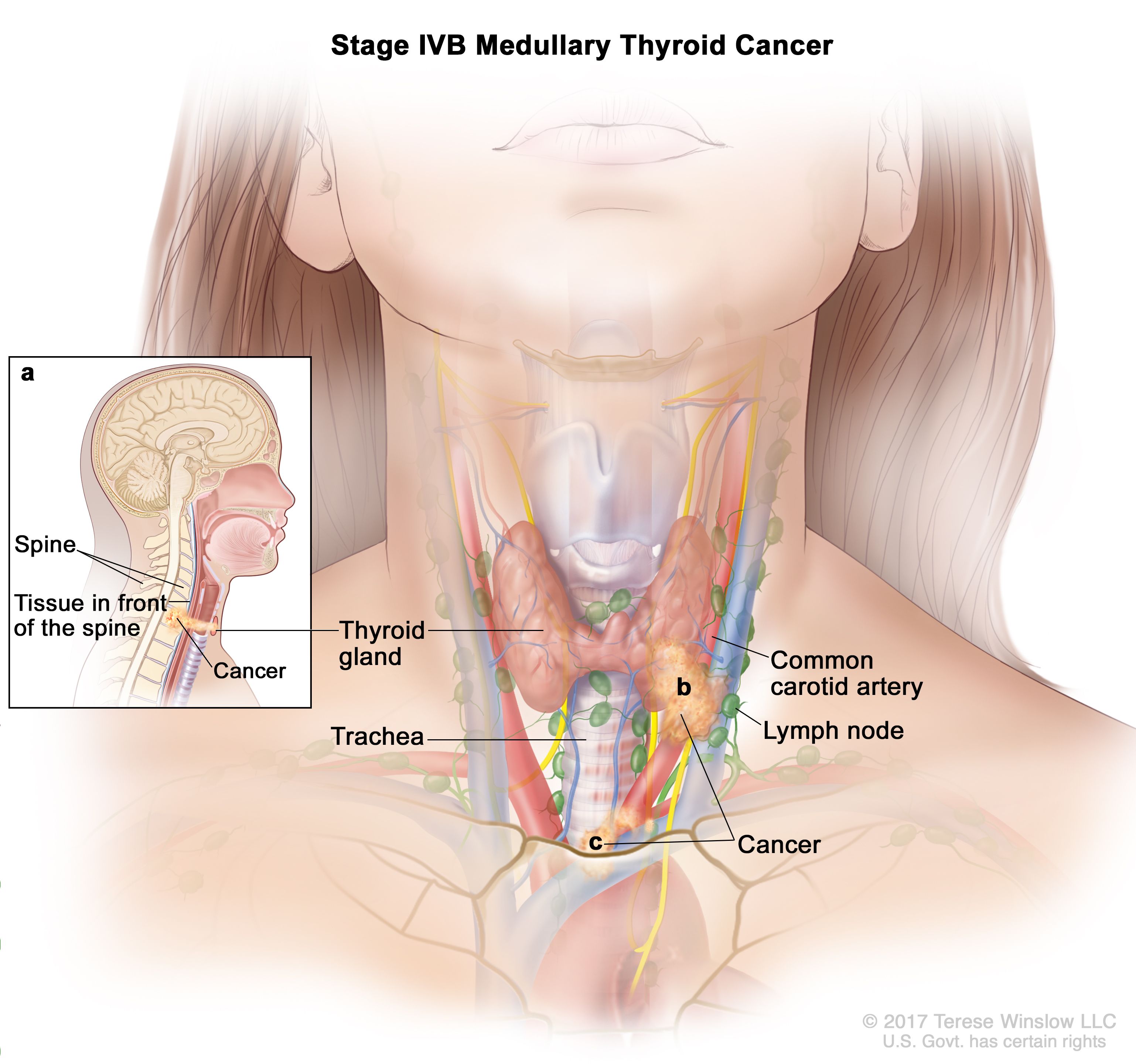
Thyroid Neck Anatomy Diagram : Medullary Thyroid Cancer Anatomy / In its anatomic position, the thyroid gland lies posterior to the sternothyroid and sternohyoid muscles.
byAdmin-
0
Thyroid Neck Anatomy Diagram : Medullary Thyroid Cancer Anatomy / In its anatomic position, the thyroid gland lies posterior to the sternothyroid and sternohyoid muscles.. Vascular anatomy and laryngeal innervation. The gland varies from an h to a u shape and is formed by 2 elongated lateral lobes with superior and inferior poles connected. Its hormones regulate basal metabolism, oxygen use, nutrient metabolism, the production of atp, and. The head rests on the top part of the vertebral column, with the skull joining at c1. Anteriorly, the sternohyoid and sternothyroid muscles overlie each of the lobes.
The thyroid gland lies between the c5 and t1 vertebrae and consists of two pear shaped lateral lobes, connected by an isthmus that the thyroid gland is in the visceral compartment of the neck along with the trachea, oesophagus and pharynx, contained within the pretracheal fascia. The thyroid is a very vascular organ and is surrounded by a sheath. Indications and complications ► the prof: Endocrine gland normally situated in the lower part of the front of the neck; Two types of hormones are produced, which are the iodine containing hormones;
Structure Of Thyroid Gland 12 Download Scientific Diagram from www.researchgate.net Download the thyroid status examination pdf osce checklist, or use our interactive osce checklist. The thyroid gland lies between the c5 and t1 vertebrae and consists of two pear shaped lateral lobes, connected by an isthmus that the thyroid gland is in the visceral compartment of the neck along with the trachea, oesophagus and pharynx, contained within the pretracheal fascia. Endocrine gland normally situated in the lower part of the front of the neck; The infrahyoid neck is the region of the neck extending from the hyoid bone to the thoracic inlet. The thyroid gland is located in the neck where it wraps around the trachea. This article describes the anatomy of the head and neck of the human body, including the brain, bones, muscles, blood vessels, nerves, glands, nose, mouth, teeth, tongue, and throat. This page is about neck anatomy thyroid cartilage,contains thyroid cartilage anatomy,functions of the larynx communication science and flashcards anatomy 2 notes hyoid bone thyroid cartilage. The effects of the hormones it produces can be seen throughout all systems in.
The thyroid needs iodine to make thyroid hormones t4 thyroxine and t3 triiodothyronine.
The gland varies from an h to a u shape and is formed by 2 elongated lateral lobes with superior and inferior poles connected. The thyroid gland lies in the neck, in front of the upper part of the trachea. This sheath attaches the thyroid to the larynx and the trachea. Anatomy of thyroid lymphatics explains patterns of spread of thyroid cancer. The head rests on the top part of the vertebral column, with the skull joining at c1. Discuss the synthesis of triiodothyronine and thyroxine. It consists of two lobes (left and right), which are connected by a central isthmus anatomical relations. The infrahyoid neck is the region of the neck extending from the hyoid bone to the thoracic inlet. A pyramidal lobe is also often present and it projects upwards from the isthmus as seen in the. It is important to know the anatomy and boundaries of the anatomical triangles of the neck. Start studying thyroid/neck anatomy & physiology. Endocrine gland normally situated in the lower part of the front of the neck; The thyroid gland is divided into two lobes that are connected by the isthmus, which crosses the midline of the upper trachea at the second and third tracheal rings.
Thyroid hormone controls metabolism and affects heart rate growth and body temperature. Anteriorly, the sternohyoid and sternothyroid muscles overlie each of the lobes. Abundant network of intraglandular lymphatics freely anastomosing between lateral lobes through the isthmus is responsible for the intrathyroidal spread. After exposing the gland through neck surgery with skin incision, the straps muscles. Endocrine gland normally situated in the lower part of the front of the neck;
Department Of Surgery Thyroid Cancer from nci-media.cancer.gov Discuss the synthesis of triiodothyronine and thyroxine. Thyroid anatomy, thyroid physiology, thyroid, thyroid gland. Learn vocabulary, terms and more with flashcards, games and other study tools. Phonation communication sciences and disorders 375 with jakielski at augustana college. Describe the location and anatomy of the thyroid gland. The head rests on the top part of the vertebral column, with the skull joining at c1. The thyroid gland is divided into two lobes that are connected by the isthmus, which crosses the midline of the upper trachea at the second and third tracheal rings. It contains blood vessels and nerves that supply structures in the head to the body.
Download the thyroid status examination pdf osce checklist, or use our interactive osce checklist.
These in humans include part of the esophagus, the larynx, trachea, and thyroid gland, major blood vessels including. The thyroid gland is closely associated with numerous other structures in the anterior neck Vascular anatomy and laryngeal innervation. Anteriorly, the sternohyoid and sternothyroid muscles overlie each of the lobes. Webmd's thyroid anatomy page provides a detailed image of the thyroid as well as a definition and information related to the thyroid. After exposing the gland through neck surgery with skin incision, the straps muscles. The thyroid gland lies in the neck, in front of the upper part of the trachea. The gland varies from an h to a u shape and is formed by 2 elongated lateral lobes with superior and inferior poles connected. Secretes, stores and releases the iodine dependent thyroid hormones thyroxine and triiodothyroine which play major endocrine roles in regulating. Thyroid hormone controls metabolism and affects heart rate growth and body temperature. The thyroid gland is an endocrine organ located in the neck that participates in a myriad of systemic processes. The neck is the part of the body, on many vertebrates, that separates the head from the torso. Your thyroid lies below your adam's apple, along the front of the windpipe.
Therefore this must be pathology arising in the visceral space. This article covers the anatomy of the thyroid gland, including functions, hormones, and clinical aspects. Describe the location and anatomy of the thyroid gland. The neck is the part of the body, on many vertebrates, that separates the head from the torso. Instant anatomy is a specialised web site for you to learn all about human anatomy of the body with diagrams, podcasts and revision questions.
Department Of Surgery Thyroid Cancer from nci-media.cancer.gov Anatomy neck vessel anatomy thyroid cancer neck lymph nodes lateral neck anatomy thyroidectomy anatomy human neck anatomy diagram organs thyroid surface anatomy normal neck anatomy thyroid anatomy and physiology strap muscles neck anatomy posterior. Ø lobes are oval shaped with rounded superior pole and elongated inferior pole. The effects of the hormones it produces can be seen throughout all systems in. Thyroid gland anatomy diagram vector illustration of thyroid gland stock vector colourbox anatomy endocrinesurgery net au The thyroid gland is a single midline endocrine organ in the anterior neck responsible for thyroid hormone production which lies in the visceral space completely enveloped by pretracheal fascia (middle layer of the deep cervical fascia). The thyroid gland is divided into two lobes that are connected by the isthmus, which crosses the midline of the upper trachea at the second and third tracheal rings. The neck is the part of the body, on many vertebrates, that separates the head from the torso. Indications and complications ► the prof:
This page is about neck anatomy thyroid cartilage,contains thyroid cartilage anatomy,functions of the larynx communication science and flashcards anatomy 2 notes hyoid bone thyroid cartilage.
The effects of the hormones it produces can be seen throughout all systems in. The head rests on the top part of the vertebral column, with the skull joining at c1. Traditionally the anatomy of the infrahyoid neck has been subdivided the swelling is centered within the borders of the thyroid cartilage. The protrusion it creates visible on the front of the neck is generally. Vascular anatomy and laryngeal innervation. The thyroid needs iodine to make thyroid hormones t4 thyroxine and t3 triiodothyronine. Webmd's thyroid anatomy page provides a detailed image of the thyroid as well as a definition and information related to the thyroid. Two types of hormones are produced, which are the iodine containing hormones; It contains blood vessels and nerves that supply structures in the head to the body. In its anatomic position, the thyroid gland lies posterior to the sternothyroid and sternohyoid muscles. Its hormones regulate basal metabolism, oxygen use, nutrient metabolism, the production of atp, and. Learn vocabulary, terms and more with flashcards, games and other study tools. The thyroid gland lies between the c5 and t1 vertebrae and consists of two pear shaped lateral lobes, connected by an isthmus that the thyroid gland is in the visceral compartment of the neck along with the trachea, oesophagus and pharynx, contained within the pretracheal fascia.
The thyroid is a very vascular organ and is surrounded by a sheath neck anatomy diagram. The gland varies from an h to a u shape and is formed by 2 elongated lateral lobes with superior and inferior poles connected.